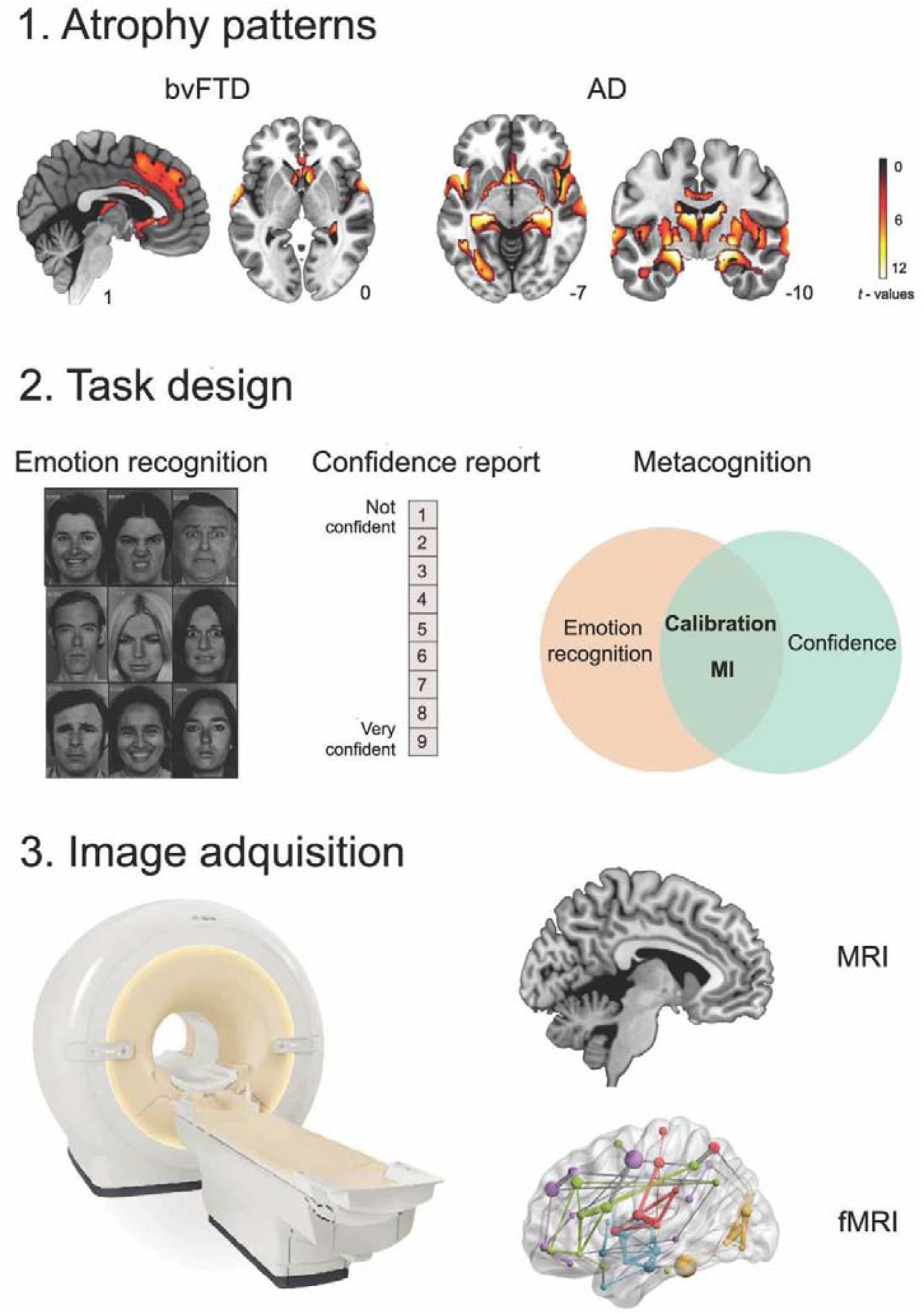
Figure 1. Patients’ atrophy patterns and data collection pipeline.

1. Atrophy of bvFTD and AD patients compared to matched controls. Neurodegeneration was observed in frontal and temporal areas for bvFTD and in posterior (temporal) regions for AD (p < 0.001, extent threshold = 50 voxels). 2. Both patient and control groups performed the emotion recognition task (Ekman’s faces) and reported their confidence about their performance. Scores of emotion recognition (accuracy) and confidence were obtained. Schematic representation of metacognition and its components: calibration (how well confidence tracks accuracy) and the metacognitive index (MI), indexing how large the difference between confidence and accuracy is. 3. Structural and functional MRI were acquired to explore the structural and functional brain correlates of emotion recognition and the MI in each patient group.
