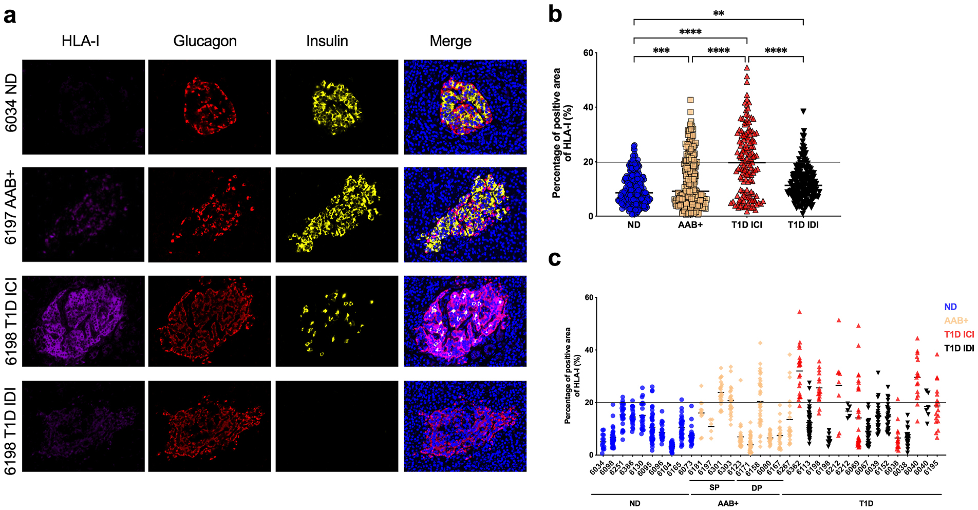
Fig. 1.

HLA class I is hyper-expressed in the islets of T1D as well as in AAB+ donors. FFPE sections of human pancreata were stained with anti-HLA-I (magenta), anti-insulin (green) and anti-glucagon (red) and Hoechst (blue). (a) Representative images of an islet from a non-diabetic (ND) donor (nPOD 6034), an autoantibody positive (AAB+) donor (nPOD 6197), an insulin-containing islet (ICI) of a T1D donor (nPOD 6198) and an insulin-deficient islet (IDI) of a T1D donor (nPOD 6198) are shown. Images were acquired using a confocal microscope LSM 780 with a 63× 1.4na objective. HLA-I was quantified using Zen software. (b) The percentage of total islet area positive for HLA-I was measured in at least 20 islets per section from non-diabetic donors (n=10), single autoantibody positive (n=5), double autoantibody positive (n=5) and type 1 diabetic donors (n=12). (c) Percentage of total islet area positive for HLA-I presented by case. Each data point represents an islet. An arbitrary threshold of 20% was set above which HLA-I was considered hyper-expressed. *** p=0.0004 ****p<0.0001.
