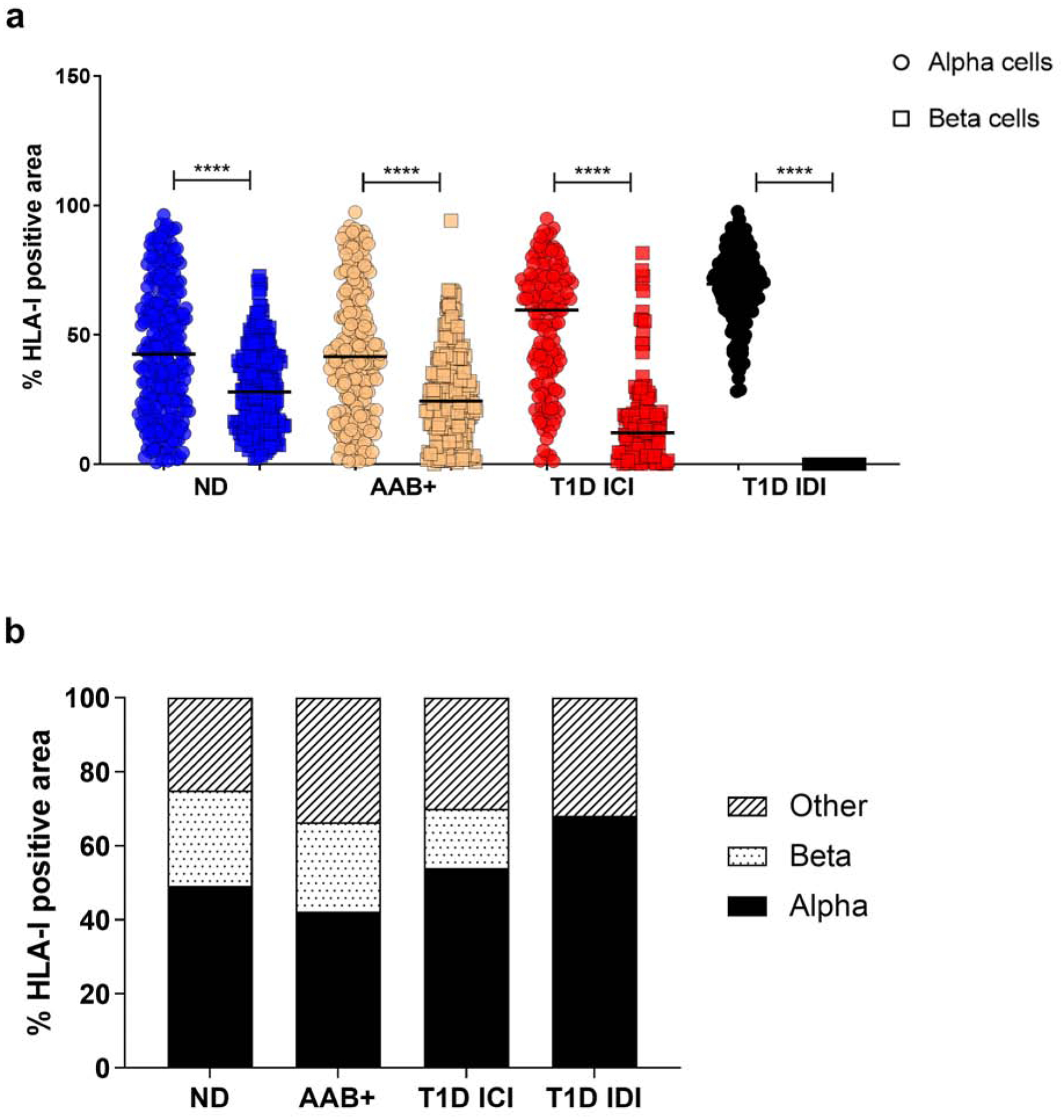
Fig. 2.

HLA class I is mainly expressed on alpha cells irrespective of disease status. (a) Percentage of colocalization of HLA-I and glucagon (alpha cells- circle) and HLA-I and insulin (beta cells - square) quantified using the Mander’s overlap coefficient using Zen software. Each data point represents an islet. (b) Mean percentage of HLA-I positive area per cell type. ND: non-diabetic; AAB+: autoantibody positive; T1D: type 1 diabetes; ICI: insulin-containing islet; IDI: insulin-deficient islet. **** p<0.0001.
