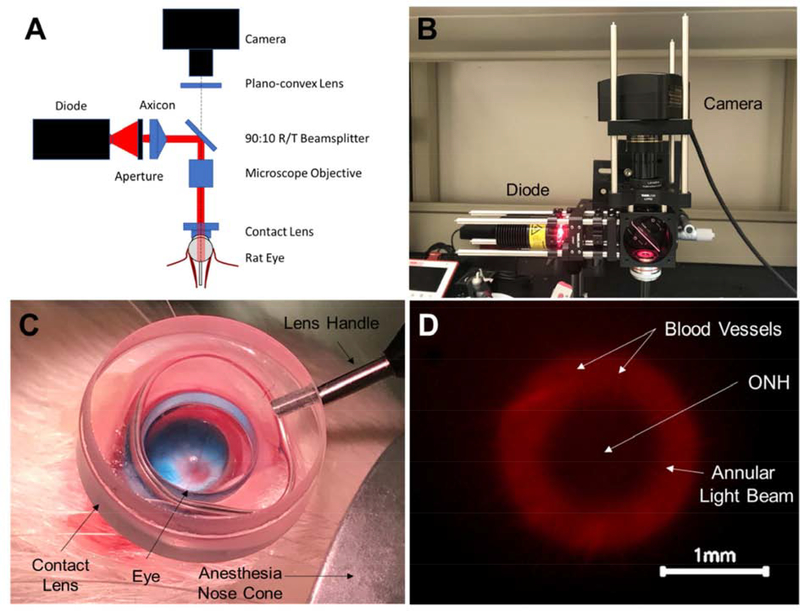
Figure 1:
Ophthalmic microscope designed to enable simultaneous transpupillary fundus visualization and annular beam projection in the rat eye in vivo. A) Component diagram, and B) photographic image of the microscope. The microscope allowed a user to simultaneously project an annular beam onto a rat fundus, observe the location of the beam on the fundus, and precisely position the beam using a three-axis manual micro-positioner stage (behind the microscope). C) A contact lens placed on the corneal surface created a clear visual path for viewing the rat fundus. The optic disc can be seen (red disc) from which blood vessels emanate. The blue color is due to the retrobulbar injection of MB, visible in this case in a non-pigmented Wistar rat. D) Image from microscope camera during illumination procedure in a Brown Norway rat (pigmented) showing annular beam spot relative to the ONH (center). The beam illumination is shown in red, with black indicating non-illuminated regions.

