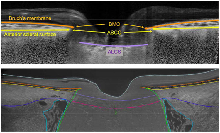
Figure 1.
Custom software to delineate ONH anatomy of the same brain-dead organ donor eye in both in vivo (before organ recovery by optical coherence tomography; OCT) and ex vivo conditions (after 3D reconstruction with episcopic autofluorescence imaging). In the OCT B-scan (upper plot), The Bruch’s membrane and opening (BMO) are marked in orange; anterior scleral surface and opening (ASCO) is marked in yellow; anterior lamina cribrosa surface (ALCS) is marked in fuchsia. In the digital section of the 3D histology reconstruction representing the same location in the ONH depicted in the OCT B-scan above. In the histology, additional landmarks of the retina are visible and delineated: the posterior lamina cribrosa surface is marked in red; pia mater is marked in green; posterior scleral surface in blue; internal surface of the dura mater in cyan. Internal limiting membrane marked in gray.

