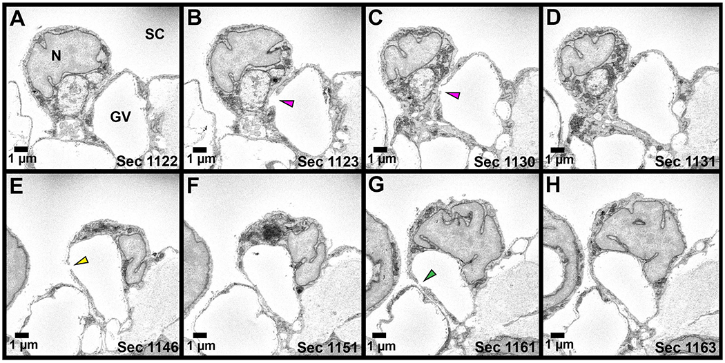
Figure 6. Serial electron micrographs through a giant vacuole (GV) with three pores.

Series of electron micrographs through a GV with three I-pores is shown. A: Section before the I-Pore 1 is observed. B-C: I-Pore 1 (pink arrowhead) spans 8 sections. D: I- Pore 1 has closed. E: I-Pore 2 (yellow arrowhead) appears sixteen sections after Pore 1 and spans 2 sections. F: I-Pore 2 has closed. G: I-Pore 3 (green arrowhead) appears fourteen sections after I-Pore 2 and spans two sections. All three I-pores appear on the same side of the GV. N = inner wall endothelial cell nucleus; SC = Schlemm’s canal.
