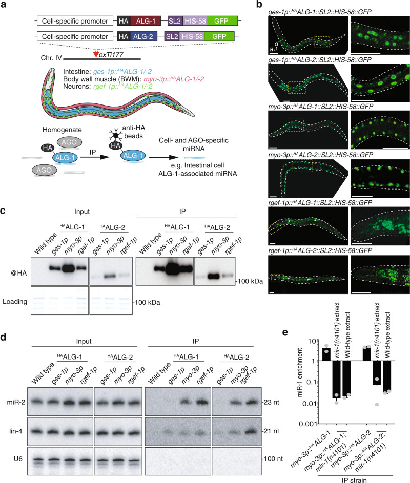
Fig. 1. Cell-type-specific profiling of AGO-loaded miRNAs.
a Overview of cell-type-specific miRNA profiling technique. HA-tagged copies of ALG-1 or ALG-2 were driven by cell-type-specific promoters, allowing immunoprecipitation of AGO-loaded miRNAs from individual tissue types from total worm homogenates. b Whole animal fluorescence images of cell-type-specific HAALG-1 and HAALG-2 lines demonstrating specific expression from intestine, body wall muscles, or neurons. Scale bars, 50 µm. Asterisks indicate intestine autofluorescence. Images are representative of >50 independent animals. c Western blot analysis of input (left) and immunoprecipitated (right) AGO complexes. All images are from the same blot and are equally exposed. Coomassie blue serves as a loading control. d RNA gel blot analysis of input (left) and AGO-immunoprecipitated miRNAs (right). U6 serves as a loading control. e Quantitative RT-PCR on immunoprecipitated AGO:miRNA complexes showing either enrichment >1 or lack of enrichment <1 of miR-1 immunoprecipitated with HAALG1 or HAALG2 in the genetic backgrounds and conditions indicated. Error bars represent +/− s.e.m. of three biological replicates. P values were calculated using one-way ANOVA with Dunnett’s multiple comparisons test. All immunoprecipitation experiments were repeated three times with similar results. HA hemagglutinin, SL2 splice leader sequence, GFP green fluorescent protein, IP immunoprecipitation.

