Fig. 3.
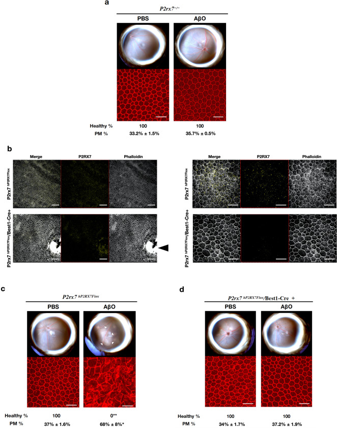
P2RX7 expression is required for AβOs-induced RPE degeneration. Eyes were treated with a single subretinal injection of 1 μM AβOs. Tissue was collected 7 days after injection. a P2rx7−/− mice are protected from AβOs-induced RPE degeneration, n = 8. b Lower magnification (left panel) and higher magnification (right panel) of RPE flat mounts of P2rx7hP2RX7Flox and P2rx7hP2RX7Flox/Best1-Cre+ mice stained with phalloidin (white) and P2RX7 (yellow) demonstrating reduction of P2RX7 signal in the RPE of P2rx7hP2RX7Flox/Best1-Cre+ mice compared to P2rx7hP2RX7Flox mice. Black arrowhead points to the optic nerve of P2rx7hP2RX7Flox/Best1-Cre+ mice, where expression of P2RX persists in non-RPE tissue. c AβOs induced degeneration in P2rx7hP2RX7Flox (n = 6) but not in P2rx7hP2RX7Flox/Best1-Cre+ mice (n = 8) (d). Representative images are shown. Fundus photographs, top row; Flat mounts stained for zonula occludens-1 (ZO-1; red), bottom row. Degeneration outlined by white arrowheads. Binary (Healthy %) and morphometric (PM, polymegethism (mean (SEM)) quantification of RPE degeneration is shown (Fisher’s exact test for binary; two-tailed t-test for morphometry; *P < 0.01, **P < 0.001). Loss of regular hexagonal cellular boundaries in ZO-1 stained flat mounts is indicative of degenerated RPE. Scale bars for lower magnification (100 μm) and higher magnification (50 μm)

