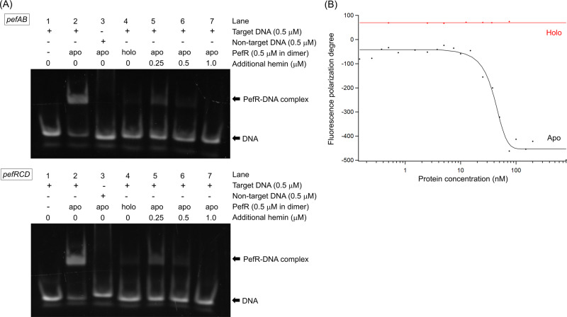
Fig. 1. DNA-binding properties of PefR.
A EMSA assays of DNA binding with/without hemin addition. The target DNAs were designed for the pefAB (top panel) and pefRCD (bottom panel) operons. The samples were prepared by mixing PefR protein with double-stranded DNA solution [20 mM Tris-HCl (pH 7.4), 50 mM KCl, 5% glycerol, and 0.2 M MgCl2], after which hemin was added to the mixture (lanes 5–7). B Fluorescence polarization assays with apo-PefR (black) and holo-PefR (red). The black line for apo-PefR is the calculated curve, giving a Kd of 40 nM, assuming a 1:1 binding ratio of the complex of apo-PefR dimer and one double-stranded DNA.

