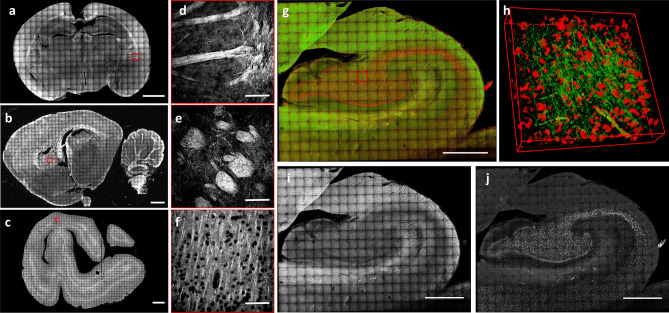
Figure 3.
High-resolution reconstruction. (a–c) Maximum intensity projection (MIP) of the reconstruction of 60-µm-thick brain sections treated with MAGIC: mouse (a), rat (b), and vervet monkey (c), respectively. Scale bar = 1 mm. (d–f) Magnified inset corresponding respectively to the red boxes in a, b, c. Scale bar = 50 μm. (g) MIP of the mesoscale reconstruction of a human hippocampus 60-µm-thick coronal section. Scale bar = 1 mm. (h) 3D rendering (450 × 450 × 60 µm3) of the stack indicated by the red box in g. (i) Green channel showing the myelinated fibers enhanced by MAGIC. (j) The red channel of the MIP in g showing the autofluorescence of the cell bodies produced by lipofuscin pigments. Images and the 3D rendering were prepared using Fiji (www.fiji.sc/Fiji)20.

