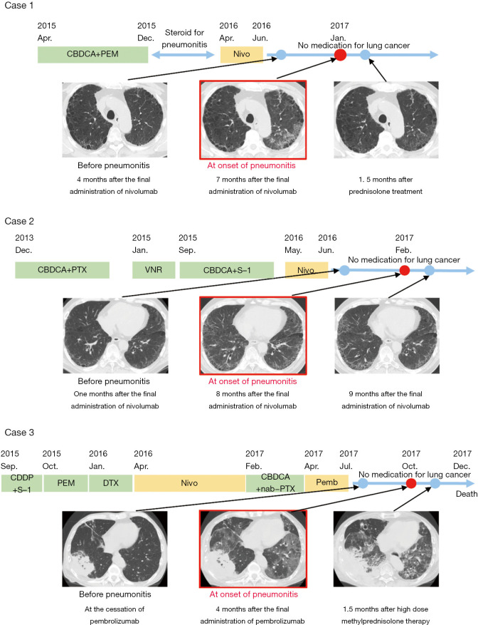
Figure 1.
Clinical course of drugs administered for lung cancer and CT images. Representative images before the onset of pneumonitis, at the diagnosis of pneumonitis and after the resolution of the pneumonitis are presented. In case 1, ground-glass opacities in the left lung improved after prednisolone treatment (image on the right, as compared to that in the middle). In case 2, slight ground-glass opacities in both lung fields improved without any medication (image on the right, as compared to that in the middle). In case 3, ground-glass opacities in both lung fields failed to improve even after high-dose methylprednisolone treatment, with contraction of the lung volume (image on the right, as compared to that in the middle).

