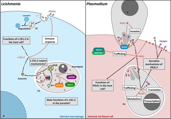
Figure 3.
Schematic representation of the pathways regulated by L-CK1.2 and PfCK1. Left panel. L-CK1.2 is localised in the parasite at the flagellar tip, the flagellar pocket, the basal body, the nucleolus and the cytoplasm as shown in brown. It phosphorylates three heat shock proteins as symbolized by brown arrows. L-CK1.2 has been identified in exosomes although the mechanisms leading to its loading into exosomes and its release are currently unknown. L-CK1.2 has functions in the macrophage, particularly in innate immunity (white box), however, most of its functions remain to be identified (grey box). Right panel. PfCK1 seems to be important for invasion either through phosphorylation of Ron3 or release by micronemes. In the parasite, PfCK1 seems to be involved in functions such as translation or trafficking (white box) but the specific mechanisms need to be elucidated. PfCK1 seems to be also released in red blood cells (RBC) as well as in the extracellular environment by unknown mechanisms. One function of PfCK1 in RBCs could be related to the regulation of trafficking through the binding of two host proteins. White boxes correspond to potential functions; Grey boxes correspond to key questions; Brown arrows correspond to known mechanisms. Black arrows correspond to mechanisms that need to be identified. F, Flagellum; FP, Flagellar pocket; BB, Basal body; k, kinetoplast; EE, Early endosome; MVB, Multivesicular bodies; N, Nucleus; CCP, Clathrin coated pit; P, phosphorylation; R, Rhoptry; M, Micronemes.

