Figure 1.
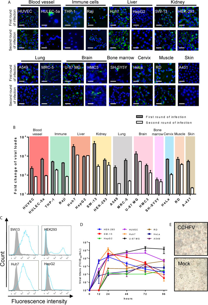
Human cell line susceptibility to CCHFV. (A) Human cell line susceptibility to CCHFV as determined by immunofluorescence assay (IFA). Cells were infected with CCHFV at an MOI of 0.01 and CCHFV nucleoprotein (NP) expression was detected at 4 days post-infection (d p.i.) using IFA. Infected cells exhibited green fluorescence (NP). Bars, 30 μm. (B) Human cell line susceptibility to CCHFV as defined by fold change of viral loads. Various cell lines were infected with CCHFV at an MOI of 0.01. Supernatants were harvested at 4 d p.i. and used for qRT-PCR. All experiments were performed in duplicate. Fold-change of viral loads were normalized to baseline viral load. (C) Flow cytometric analysis of CCHFV-infected cells. Different cell lines were infected with CCHFV at an MOI of 0.01. Cells were harvested at 2 d p.i. and expression of CCHFV NP was detected using fluorescein isothiocyanate (FITC)-conjugated CCHFV NP monoclonal antibody and analyzed using flow cytometry. The shaded area represents the control. The mean florescence intensity (MFI) of mock-infected/CCHFV-infected were 837/10200, 44/6491, 179/5528 and 375/839 for SW13, HEK293, Huh7 and HepG2 cells respectively. At least 10000 cells were used for analysis. (D) Viral characteristics of CCHFV in 6 human cell lines. The different cell lines were infected with CCHFV at an MOI of 0.01. Supernatants were harvested at the indicated time points post infection. Viral titers in the supernatants were determined using end-point dilution assays. Means ± SD of three represented independent experiments are shown. (E) SW-13 cells were infected with CCHFV at an MOI of 0.01. Cytopathic effects of cell rounding and detachment were evaluated at 4 d p.i. using an inverted microscopy. Mock, uninfected SW-13 control.

