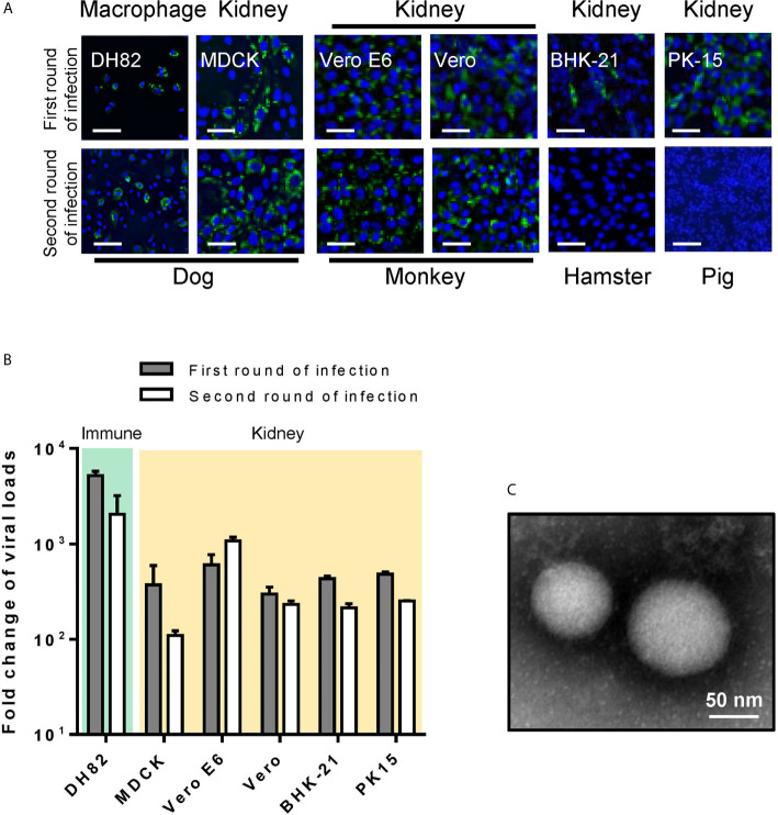
Figure 2.
Animal cell line susceptibility to CCHFV. (A) Animal cell line susceptibility to CCHFV was determined by IFA. Cells were infected with CCHFV at an MOI of 0.01 and CCHFV NP expression was detected at 4 d p.i. using IFA. Infected cells exhibited green fluorescence (NP). Bars, 30 μm. (B) Animal cell line susceptibility to CCHFV as defined by fold-change of viral loads. The different cell lines were infected with CCHFV at an MOI of 0.01. Supernatants were harvested at 4 d p.i. and evaluated using qRT-PCR. All experiments were performed in duplicate. Fold-change of viral loads were normalized to baseline viral load. (C) Electron micrographs of negative staining of CCHFV particles in cell culture supernatants from Vero E6. Bar, 50 nm.

