
Fig. 3.
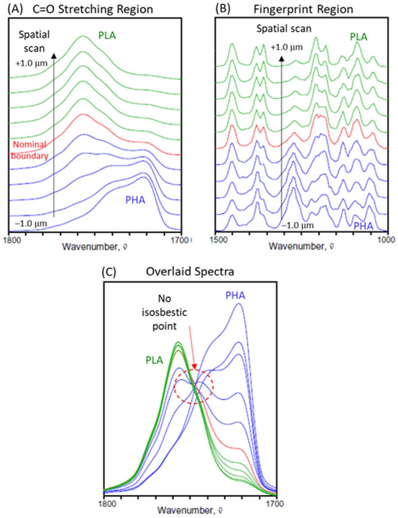
O-PTIR spectra of the laminate cross section collected every 200 nm around the PHA/PLA interface in the C=O stretching (A) and finger print region (B) regions, as well as the overlaid plot of spectra in the C=O stretching region (C).
Official websites use .gov
A
.gov website belongs to an official
government organization in the United States.
Secure .gov websites use HTTPS
A lock (
) or https:// means you've safely
connected to the .gov website. Share sensitive
information only on official, secure websites.
O-PTIR spectra of the laminate cross section collected every 200 nm around the PHA/PLA interface in the C=O stretching (A) and finger print region (B) regions, as well as the overlaid plot of spectra in the C=O stretching region (C).