Fig. 3.

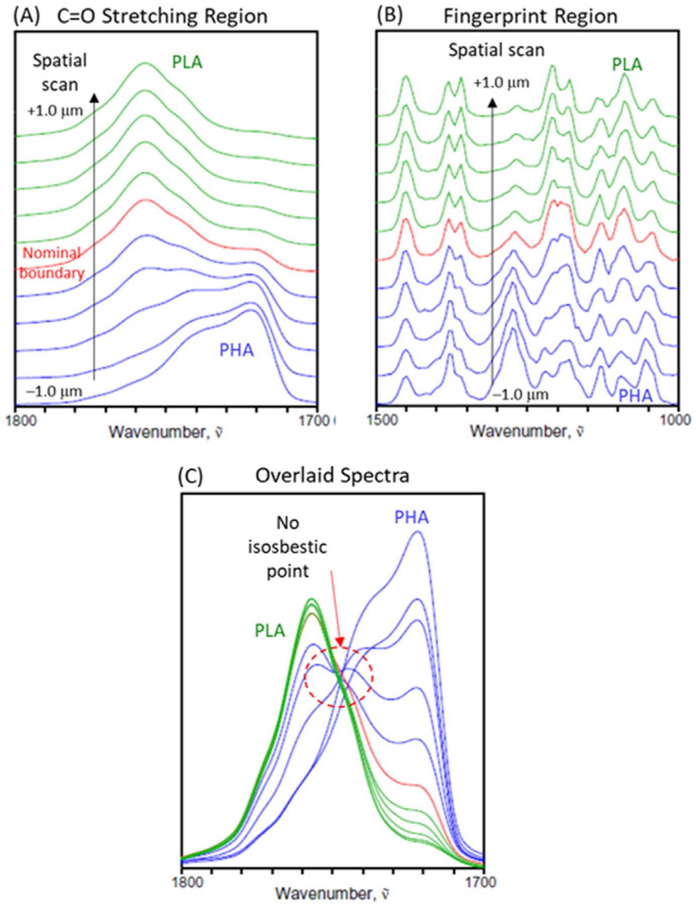
O-PTIR spectra of the laminate cross section collected every 200 nm around the PHA/PLA interface in the C=O stretching (A) and finger print region (B) regions, as well as the overlaid plot of spectra in the C=O stretching region (C).

O-PTIR spectra of the laminate cross section collected every 200 nm around the PHA/PLA interface in the C=O stretching (A) and finger print region (B) regions, as well as the overlaid plot of spectra in the C=O stretching region (C).