
Figure 5. Induction of micronuclei by luteolin in TK6 cells transduced with EV and various CYPs.
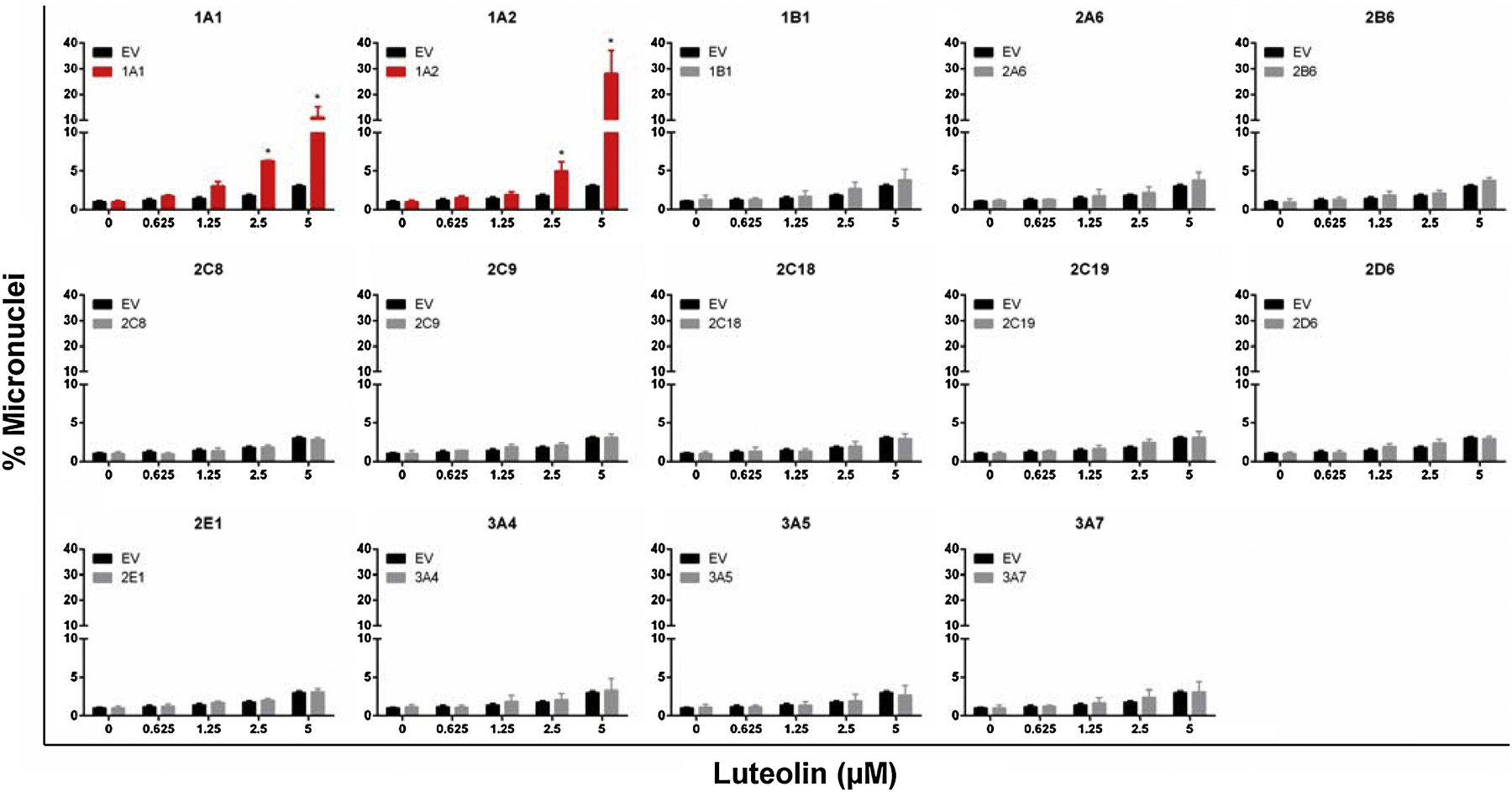
A high-throughput micronucleus assay was used to quantify the micronuclei. The stopping gate was set to record 10,000 intact nuclei. The data represent mean ± SD from at least three independent experiments. * indicates P < 0.05 comparing a CYP-expressing cell line versus the corresponding EV cells at the same concentration.