
Figure 9. Effects of diosmetin on cytotoxicity and genotoxicity in different TK6 cell lines.
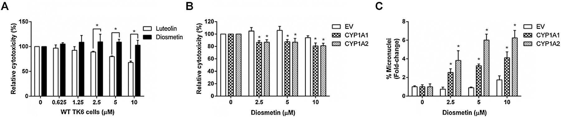
Cells were treated with diosmetin at the indicated concentrations for 24 h. (A & B) Cytotoxicity was measured by the cellular ATP level using the CellTiter-Glo® assay. (C) Genotoxicity was determined by the high-throughput micronucleus assay using flow cytometry. The data represent as mean ± SD from three independent experiments * indicates P < 0.05 comparing diosmetin and luteolin at the same concentration (A) or a CYP-expressing cell line versus the EV controls at the same concentration (B & C).