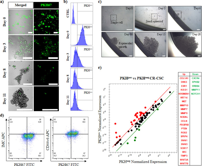
Figure 1.
Identification of quiescent colorectal cancer stem cells (PKHpos qCR-CSCs #21). Proliferative PKH67 staining assay enables to identify, after 11 days, the qCR-CSCs by (a) confocal microscopy (the green cells) and (b) flow cytometry analysis. The y axis of the histograms represents the counts, while the x axis represents the fluorescent signal measured for green PKH67. (c) Self-renewal capability of PKHpos qCR-CSC sorted by FACS and cultured in an isolated environment (one cell per well, scale bars: 50 μm). (d) Flow cytometry analysis of CD44v6 in PKH67-stained CR-CSC (#21) after 11 days of PKH67 staining. (e) Stemness-related gene expression analysis of up- and downregulated genes in FACS-sorted PKHpos and PKHneg CR-CSCs following 11 days of PKH67 staining. Genes showing a more than threefold change in PKHposg than PKHneg CR-CSCs are shown in red and green, respectively.

