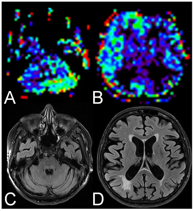
Figure 2. Example of a crossed cerebellar diaschisis-positive patient with Alzheimer’s disease with extensive reduced relative cerebral blood flow in the ipsilateral hemisphere. Arterial spin labeling perfusion magnetic resonance images showed reduced relative cerebral blood flow in the right cerebellum (A) and the left cerebral hemisphere (B). On fluid-attenuated inversion recovery images the cerebellum (C) and the left cerebral hemisphere (D) were unremarkable. Figure 1. Example of a crossed cerebellar diaschisis and ipsilateral thalamic diaschisis-positive patient with Alzheimer’s disease. Arterial spin labeling perfusion magnetic resonance images showed reduced.

