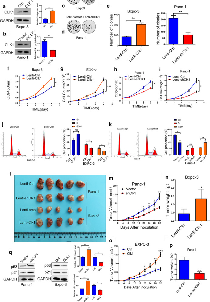
Fig. 2.
CLK1 enhanced the proliferation ability of human pancreatic cancer cells in vitro and in vivo. a, b BxPC3 cells with stable CLK1 overexpression (a) and PANC-1 cells with CLK1 knockdown (b) were generated. The changes in CLK1 expression were confirmed using western blot assays. c–i The proliferative ability of stably transfected PANC-1 or BxPC3 cells was investigated via colony formation assays (c–e) and CCK-8 assays (f–i). Representative colony formation images are shown (c, d), and the numbers of colonies were summarized (e). j, k Flow cytometry analysis of the cell cycle progression of stably transfected PANC-1 (k) or BxPC3 (j) cells was performed. Representative images and quantification of the results are presented. l–n Overexpression of CLK1 promoted the growth of BxPC-3 cells in a subcutaneous xenograft mouse model (l–n). The size of the tumors was measured at the indicated time points (m, ***P < 0.001). Tumors were extracted and weighed after mice were sacrificed (n, ***P < 0.001). CLK1 knockdown inhibited the growth of PANC-1 cells in a subcutaneous xenograft mouse model (l, o, p). The size of the tumors was measured at the indicated time points (o, ***P < 0.001). Tumors were extracted and weighed after mice were sacrificed (p, ***P < 0.001). q The protein levels of p21 and p53 were quantitated by western blot assays, and representative images are shown. Data are shown as mean ± SD from three independent experiments. *P < 0.05; **P < 0.01; ***P < 0.001, between the indicated groups

