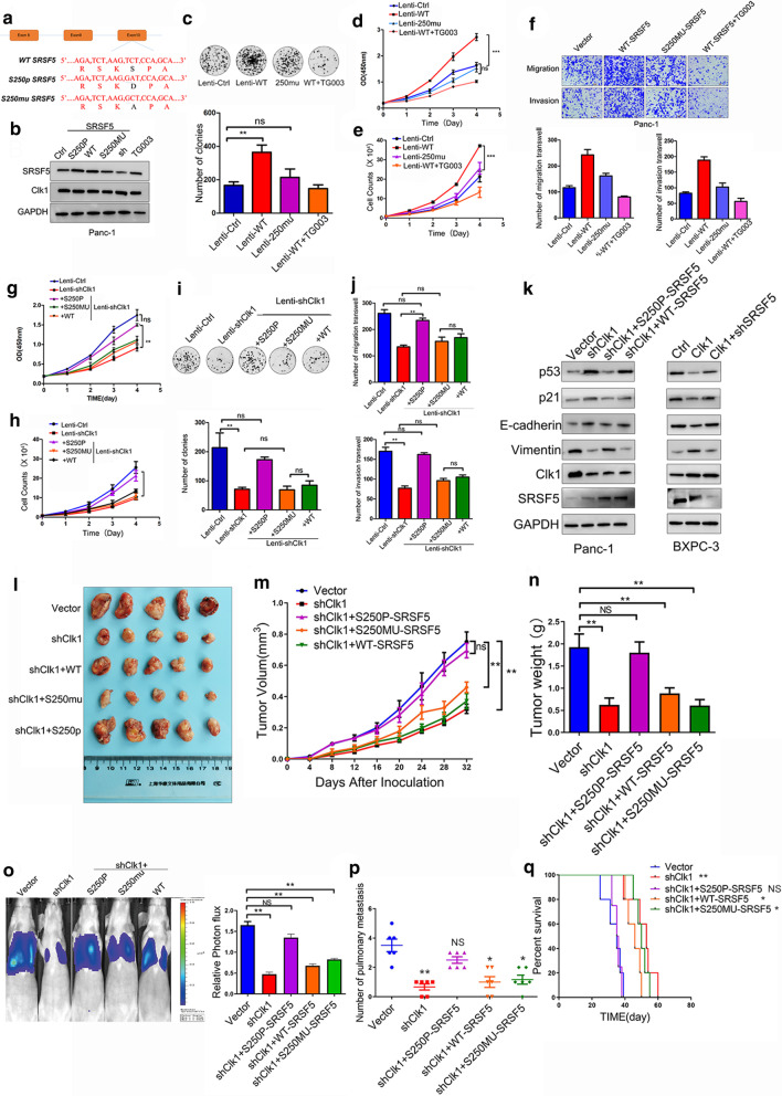
Fig. 5.
CLK1-induced SRSF5 phosphorylation on Ser250 contributed to the proliferation and metastasis of pancreatic cancer cells. a A schematic diagram shows the nuclear and amino acid sequences of wild type, mutated, and phosphorylated SRSF5 at Ser250 site. b PANC-1 cells were left untreated (Ctrl), or transduced with plasmids expressing wild type (WT)/mutated (S250MU)/phosphorylated (S250P) SRSF5, or treated with TG003 for 24 h. The protein levels of SRSF5 and CLK1 were determined by western blot assays. c–f The colony formation (c) and proliferation (d, e) ability, migration and invasion ability (f) of the indicated cell lines were evaluated. g–k PANC-1 cells were infected control lentivirus, or lentivirus expressing CLK1-specific shRNA, or lentivirus expressing CLK1 shRNA together with lentivirus expressing wild type (WT)/mutated (S250MU)/phosphorylated (S250P) SRSF5. The cell proliferation ability (g, h), colony formation ability (i), migration and invasion ability (j) of the indicated cells were determined. The protein levels of p21, p53, E-cadherin, and Vimentin in the indicated cells were determined by western blot assays (k). l–q PANC-1 cells as indicated in (h) were inoculated into nude mice, and mice were sacrificed. The images of tumors (l), bioluminescence images of mice (o), and tumor growth curves (m), tumor weight (n), mice survival (p), and number of pulmonary metastasis (q) are shown. All data are shown as mean ± SD from three independent experiments. *P < 0.05; **P < 0.01; ***P < 0.001, between the indicated groups

