
Fig. 3.
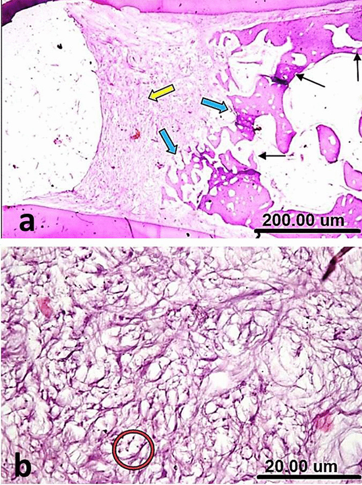
(a) A sample of group III (Positive control) after one week showing the furcation defect filled with granulation tissue, no calcific bridge formation (yellow arrow), irregular silhouette of interradicular bone at the granulation tissue interface (blue arrows) and distorted thin discontinuous trabeculae (thin black arrows). (b) A higher magnification of the same sample in Fig. 3a showing absence of prominent calcification at the furcation perforation zone and many inflammatory cells scattered among collagen fibers (red circle) [H&E, X4 (a) and X40 (b)]