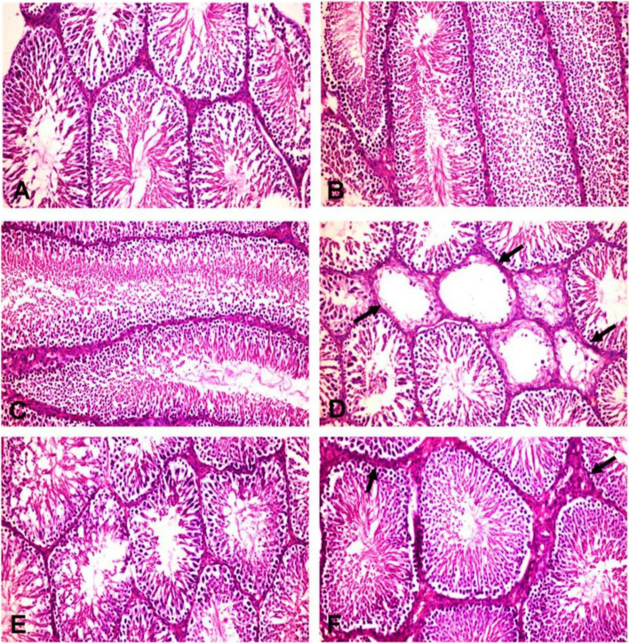
Fig. 3.
Histopathological pictures of the testes in different experimental groups (H&E X200). a Control group, b POS (200 mg/kg) and (c) POS (400 mg/kg) treated groups; showing normal histology of the seminiferous tubules with main spermatogenic series, Sertoli cells and Leydig cells. d ACR treated group showing some seminiferous tubules suffering from marked testicular degeneration (arrows) with a complete absence of spermatogenic series. e ACR + POS (200 mg/kg) treated group showing normal seminiferous tubules with incomplete spermatogenic series. f ACR + POS (400 mg/kg) treated group showing normal seminiferous tubules with thickening of intertubular tissue

