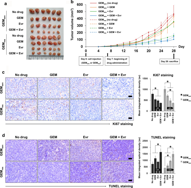
Fig. 7.
In vivo evaluation of the individual and combined effect of GEM and Evr on pancreatic tumor progression. Six-week-old BALB/C nude mice were subcutaneously injected with GEMsen or GEMres cells and administered GEM or/and Evr after one week of tumor formation. a Macroscopic observation of tumors extracted from xenografted animals at the end of the 28-day experimental period. b Tumor growth curve showing the change in tumor volume over the experimental period. Tumor volume was measured every two days. GEMsen and GEMres tumors were denoted by solid and dashed lines, respectively. c Ki67 staining and quantification of tissue proliferation in GEMsen- or GEMres-transplanted tumors treated with GEM or/and Evr. Scale bar, 50 μm. d TUNEL staining and quantification of tissue apoptosis in GEMsen- or GEMres-transplanted tumors treated with GEM or/and Evr. Scale bar, 50 μm. The relative amount of Ki67-positive and TUNEL-positive staining was evaluated using ImagePro Plus and expressed as the mean integrated optical density of brown staining. The numbers in the arrows represent the difference (increase or decrease) in positive staining between GEM and GEM + Evr. In GEM-treated GEMres tumors, the suppressive effect of Evr on tissue proliferation and the enhancing effect of Evr on tissue apoptosis were stronger than those in GEM-treated GEMsen tumors. The data are expressed as the mean ± standard deviation of six replicates (n = 6). *P < 0.05; #P < 0.05 compared with the same treatment in GEMsen tumors. GEM: gemcitabine, Evr: everolimus; GEMsen: GEM-sensitive pancreatic cancer tumors; GEMres: GEM-resistant pancreatic cancer tumors; TUNEL: terminal deoxynucleotidyl transferase dUTP nick end labeling; au: arbitrary units

