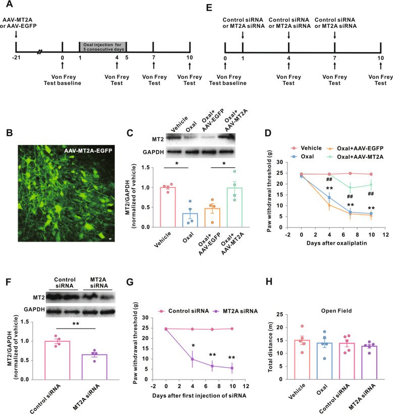
Fig. 2.
Impact of genetic modification of MT2 on Oxal-induced neuropathic pain. a–e Experimental timeline for b–d, f–h, Fig. 3, and Fig. 5. The arrows above the timeline mark the injection of AAV-EGFP-MT2A or AAV-EGFP (a) or mark the injection of MT2A siRNA or control siRNA (e); the arrows below the timeline mark the von Frey test time points. The administration of vehicle or Oxal (0.4 mg/100 g/day for five consecutive days) was carried out 21 days after intraspinal injection of AAV-MT2A-EGFP in male rats. b Neuron-specific green fluorescence in the L5 spinal dorsal horn in rats 21 days after intraspinal injection of AAV-MT2A-EGFP. Scale bar, 100 μm. c MT2 protein expression in the groups as indicated. Representative western blots (top panels) and a summary of densitometric analysis (bottom graphs). *p < 0.05; n = 4. d Hind paw withdrawal threshold response to von Frey filament stimuli on days 0, 4, 7, and 10 after the treatment. **p < 0.01 versus vehicle, ##p < 0.01 versus Oxal + AAV-EGFP; n = 8 in Oxal + AAV-EGFP; n = 11 in Oxal + AAV-MT2A-EGFP. f Intrathecal injection of MT2A siRNA reduced protein expression of MT2A. **p < 0.01; n = 4. g Hind paw withdrawal threshold in normal male rats. *p < 0.05 and **p < 0.01 versus control siRNA; n = 6. h Oxaliplatin administration or MT2A siRNA did not show reduced locomotor activity 10 days after oxaliplatin or vehicle treatment (n = 5). Oxal, oxaliplatin

