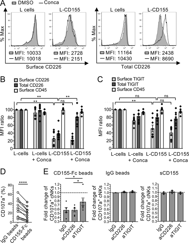
Figure 4. Membrane-bound CD155 induces CD226 degradation and NK cell dysfunction.
(A-C). cNKs isolated from MP were incubated with L cells or L-CD155 for 16 h +/− concanamycin-A (Conca). (A) Representative histograms of flow cytometry showing the effects of Conca on cell surface and total CD226 expression (MFI) by NK cells in the presence of L-CD155 cells or L cells as compared with DMSO control. (B and C) Cell surface and total CD226 (B), TIGIT (C), and surface CD45 (as control) expressions by cNKs (normalized MFI expression as compared to cNKs + L cells). (D and E) cNKs were incubated 48 h with either sCD155, IgG- or CD155-Fc-beads +/− blocking antibodies and washed before functional assay in presence of FO-I. (D) CD107a expression (%) by cNKs pre-incubated or not with CD155-Fc beads (n=11) (E) CD107a expression (fold change) by cNKs pre-incubated with either CD155-Fc-beads (n=6), IgG-beads (n=3), or sCD155 (n=3) +/− aCD226 or aTIGIT blocking mAbs as compared with IgG-beads (dotted line). Horizontal bars depict means ± SEM (B, C, and E) and P values were obtained by one-way ANOVA tests followed by Tukey’s (B and C) or Dunnett’s (E) multiple comparisons test or by paired t-tests (D) with ns (non-significant), P>0.05; *, P<0.05; **, P<0.01 and ****, P<0.0001. Data are representative of at least three independent experiments done in duplicates.

