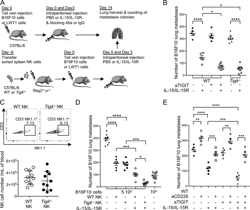
Figure 6. IL-15 and TIGIT blockade/deletion combine to suppress experimental lung metastasis of melanoma.
(A) Schematic of the mice tumor immunotherapy experiments. (B and E) Groups of C57BL/6 WT and Tigit−/− mice (n=5 mice/group) were injected i.v. with (B) 5 × 10⁵ B16F10 or (E) 2 × 10⁵ B16F10 on day 0. Some groups of mice then received PBS or IL-15 (0.5 μg)/IL-15Ra (3.0 μg) i.p., cIg or anti-TIGIT (200 μg) on days 0 and 3 and/or anti-CD226 (250 μg) on days −1, 0 and 7 (E). (C) Freshly sorted NK cells (TCRβ−NK1.1+) from spleens of wild-type (WT) or TIGIT−/− male mice were injected intravenously into Rag2−/−γc−/− recipient male mice (2 × 10⁵ cells per mouse). At day 6, peripheral blood was collected from Rag2−/−γc−/− recipient mice to check NK cell reconstitution. Data are shown as representative flow and quantitative results of NK cell reconstitution. (D) Groups of C57BL/6 Rag2−/−γc−/− (n=6–10 mice/group) were i.v. reconstituted with 2 × 10⁵ purified WT or Tigit−/− NK cells. Six days later, reconstituted mice were injected i.v. with 5 × 10⁵ or 1 × 10⁴ B16F10 (day 0). Some groups of mice then received PBS or IL-15 (0.5 μg)/IL-15Rα (3.0 μg) i.p. on d 0 and 3. In B-E on day 14, lungs were harvested, and the metastatic burden was quantified by counting colonies on the lung surface. Data are presented as mean ± SEM. Each experiment was performed once. Legends indicates the group of mice and the treatment per condition. Statistical significance was determined by one-way ANOVA with Tukey’s multiple comparisons test with *, P<0.05; **, P<0.01; ***P<0.001 and ****, P<0.0001.

