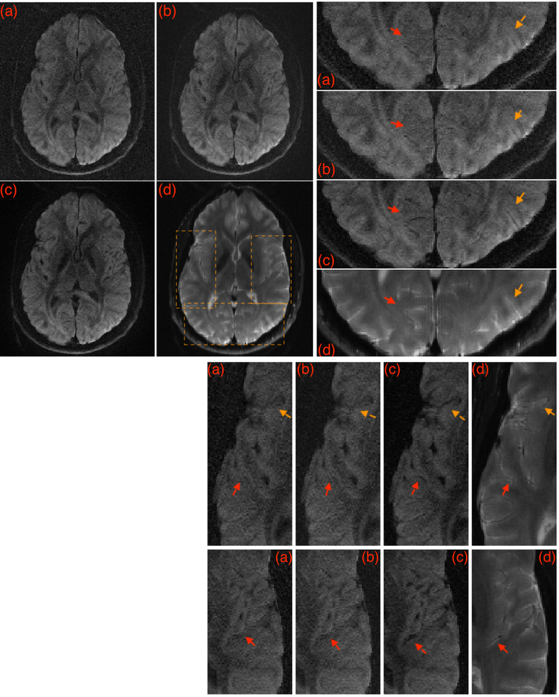
Figure 2:
A given DWI reconstructed using various methods. (a) shows MUSE reconstruction, (b) shows IRLS MUSSELS without CS, and (c) shows the IRLS MUSSELS with CS constraint. In (d) the sum-of-squares reconstruction of the b0 image from the same slice location is provided for comparison of the anatomical details from the slice, that is unaffected by the reconstruction method. The boxed regions are zoomed for better visualization. Arrows highlight regions with better recovery of anatomical details.

