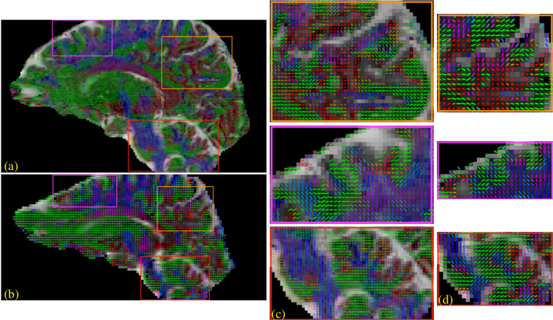
Figure 3:
Whole brain reconstruction of dataset 2. The IRLS with CS reconstruction was performed on the dataset and a single tensor model was fitted to the DWIs. This 1.1 mm isotropic dataset (a,c) is compared against the 2mm isotropic data (b,d) obtained from the same subject. Several regions are highlighted where the high-resolution data offers more details about the brain anatomy which is not fully captured by the low resolution data. (c) shows the zoomed view of regions highlighted in (a) and (d) shows the zoomed view of regions highlighted in (b).

