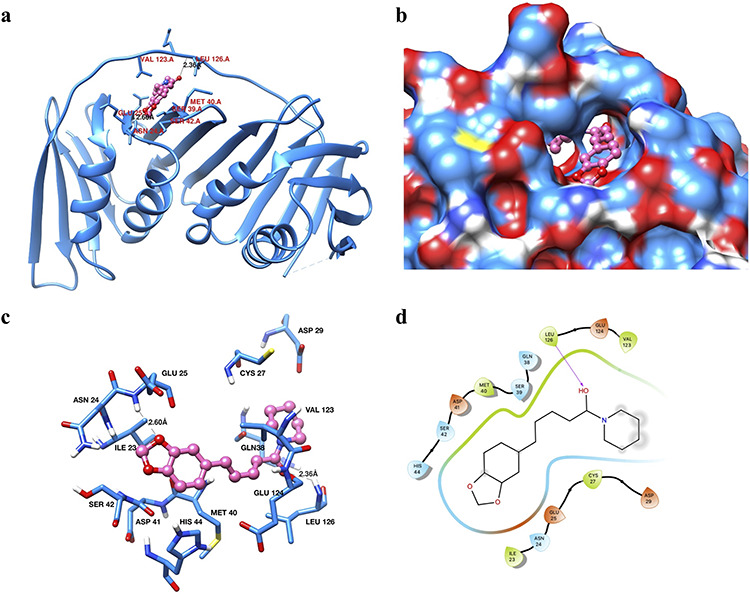
Figure 10.

binding analysis of piperine with PCNA. (a) Ribbon diagram of PCNA protein showing the different secondary structure elements and PIP in the binding site pocket (shown in hot pink color and ball and stick representation). Interacting residues are shown in dodger blue color as sticks. (b) Surface diagram of PCNA showing the active site pocket with piperine (hot pink color and ball-stick representation). (c) Interactions of piperine with the active site residues of PCNA. Interacting residues are in dodger blue color and shown in sticks representation. Piperine is shown in hot pink color as a ball and stick. Hydrogen bonds are labeled and shown as black dashed lines. (d) 2D interaction diagram of piperine with PCNA.
