
Figure 3.
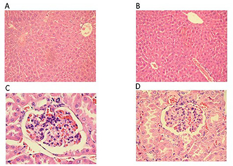
Histological sections of the liver and kidney of CD-1 mice administered orally with 3000 mg/kg MPJ daily for 120 days. Sections were stained with hematoxylin and eosin. (A) 10X liver section of control group (B) 10X liver section of group administered with 3000 mg/kg MPJ. Both sections show a normal histological structure of hepatocytes showing the central veins. (C) 40X kidney section of control group. (D) 40X kidney section of group administered with 3000 mg/kg MPJ. Both sections show normal structures for Bowmen's capsule, glomerulus, and proximal tubule.