
Figure 5.
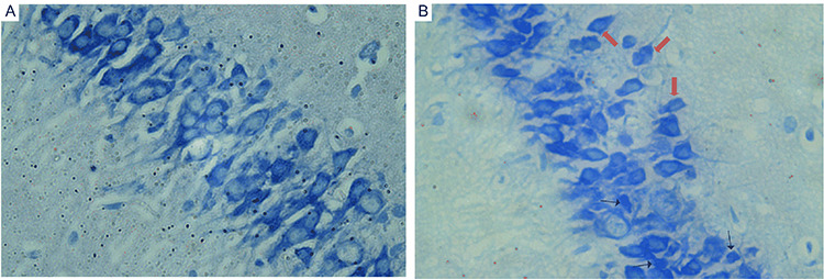
The morphological changes of coronal sections of CA1 in hippocampus were observed with light microscope after Nissl staining (magnification of ×400). (A) Control group and (B) NaAsO2 exposed group. Increased number of degenerating neurons with smaller cell bodies or pyknotic nuclei (black arrow) and ectopic pyramidal cells (red arrow) can be found in hippocampus of NaAsO2 exposedmice.