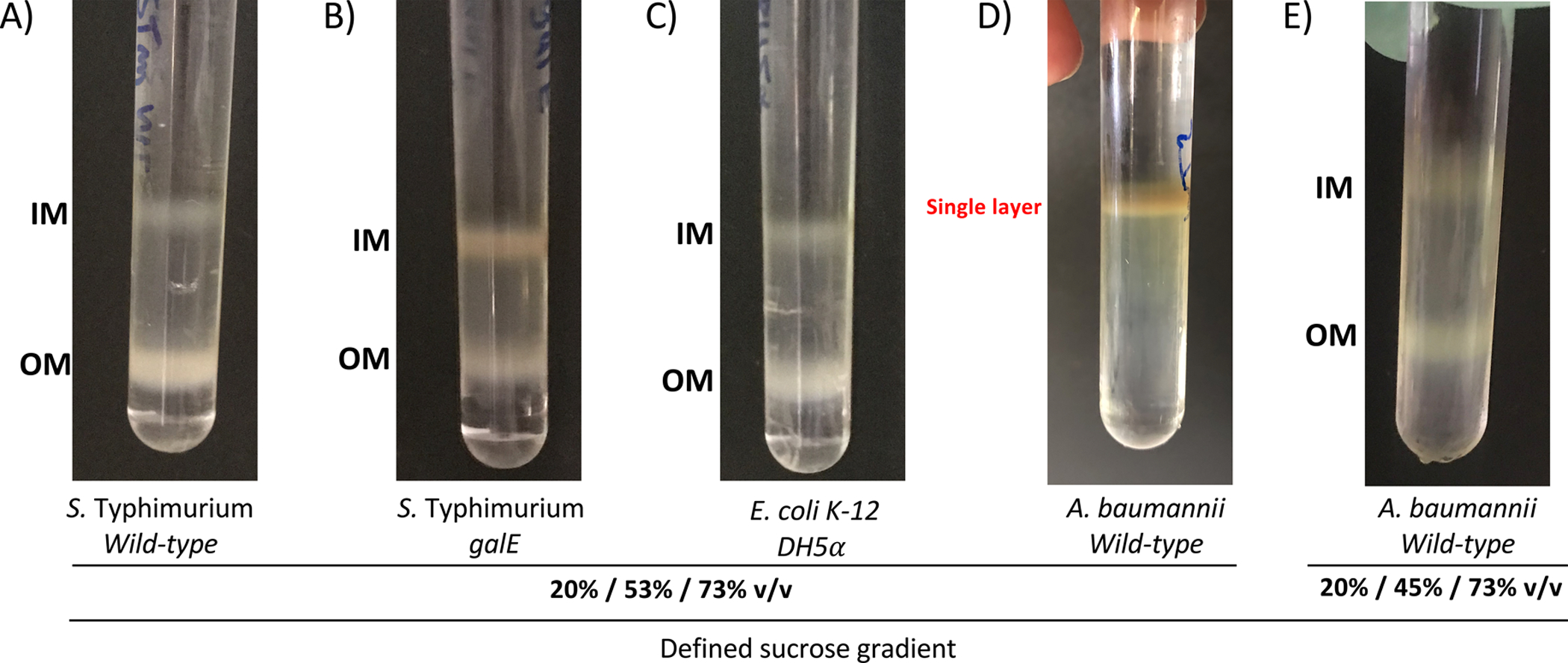
Figure 2. Representative results for different Gram-negative species whose membranes were isolated using the standard and the modified sucrose density gradients described in this article.

Images of discontinuous sucrose density gradients post isopycnic centrifugation for A. wild-type Salmonella enterica serovar Typhimurium 14028s, B. galE-mutant S. Typhimurium LT2, which produces LPS molecules that are devoid of O-antigens, and C. Escherichia coli K-12 DH5α, which also produces LPS molecules that lack O-antigens. The inner membrane (IM) is separated from the outer membrane (OM) and localizes to the 20–53% sucrose interface as a brown material. The white OM layer localizes to the 53–73% sucrose interface due to the higher density of this fraction. D. The total membranes of the wild-type Acinetobacter baumanii 17978 did not separate using 20%/53%/73% w/v sucrose gradient, E. but did separate using the 20%/45%/73% w/v sucrose gradient.
