
Figure 2.
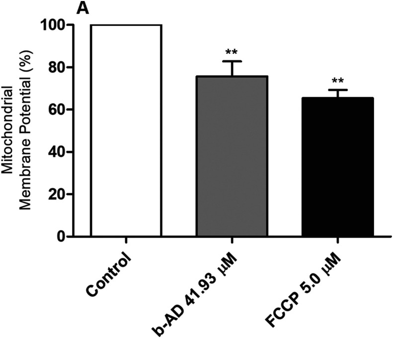
Evaluation of mitochondrial membrane potential. Leishmania infantum (107 cells) were incubated alone (control) or in the presence of b-AD (41.93 μM, corresponding to two times the IC50 value) for 24 h at 25 °C. Parasites were then incubated with MitoTracker Red CM-H2XROS for 30 min and in the dark. Cells were washed twice with PBS and transferred to a black 96-well plate, when the fluorescence intensity was evaluated using a fluorometer. Promastigotes treated with carbonyl cyanide-4-(trifluoromethoxy)phenylhydrazone (FCCP; 5.0 μM) were used as a positive control, while those untreated were used as a negative control (control). Bars represent the mean plus standard deviation of the groups. (**) indicates a statistically significant difference as compared to the control (p < 0.0001).