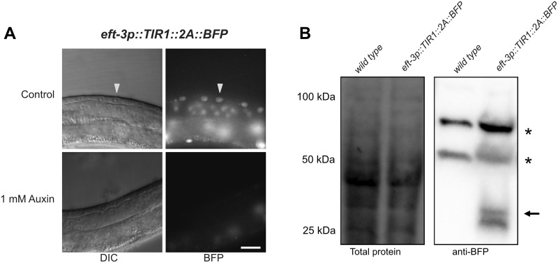
Figure 3.
The F2A ribosome skip sequence functions efficiently in an eft-3p::TIR1::F2A::BFP::AID*::NLS transgene. (A) L3 larvae expressing eft-3p:: TIR1::F2A::BFP::AID*::NLS. A control animal expresses AID*-tagged BFP in the nuclei of vulval precursor cells (VPCs; white arrows). BFP expression is undetectable in animals grown on 1 mM K-NAA—a water-soluble, synthetic auxin—for 1 hour before imaging. Scale bars represent 15 µm (eft-3p). (B) Western blot detecting BFP::AID::NLS. Stain-free (Bio-Rad) analysis of total protein on the blot is provided as a loading control (left). Marker size (in kilodaltons) is provided. Anti-BFP blot showing background bands (marked with *) and a doublet consistent with the predicted size of BFP::AID*::NLS (black arrow) at approximately 34.5 kDa, and a smaller band below, likely a BFP degradation product.

