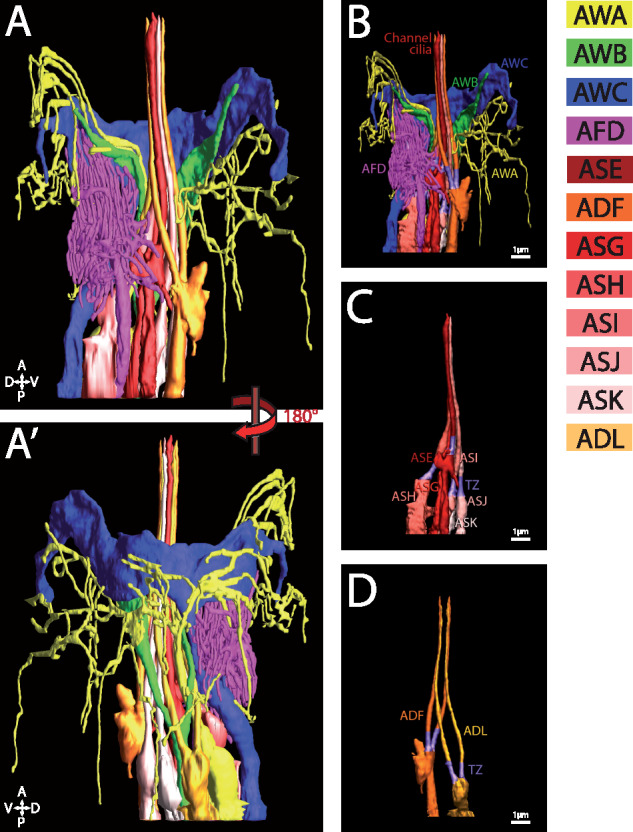
Figure 1.

Cilia of amphid sensory neurons. (A and A′) 3 D reconstruction model of the sensory endings of 12 amphid neuronal cilia on the right side. Complex sensory endings of the winged cilia of AWA, AWB, and AWC and microvilli of the AFD neurons are shown in (B). Single (ASH, ASG, ASE, ASI, ASJ, and ASK) and double rod-shaped (ADF and ADL) channel cilia are shown in (C) and (D), respectively. Individual amphid neurons are color coded as indicated. Scale bar: 1 µm. Adapted from Doroquez et al. (2014).
