Figure 4.
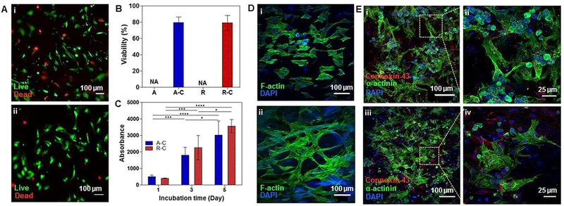
(A) Live/Dead staining of cardiac fibroblasts seeded on (i) PEDOT-embedded aligned fibers and (ii) PEDOT-embedded random fibers. (B) Quantification of Live/Dead images. (C) Metabolic activity of cardiac fibroblasts seeded on the PEDOT-embedded aligned and random fibers (n=3 in B, C). (D) F-actin and DAPI staining of cardiac fibroblasts seeded on PEDOT-embedded (i) aligned and (ii) random fibers. (E) Immunostaining of sarcomeric α-actinin and Cx-43 of cardiomyocytes seeded on (i and ii) PEDOT-embedded aligned fibers and (iii and iv) PEDOT-embedded random fibers.

