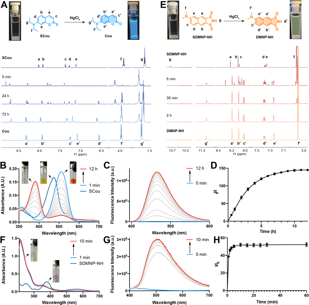
Fig. 2.
Spectral changes of SCou and SDMNP-NH after Hg2+ addition. A) 1H NMR monitoring of SCou 1 mM with 10 eq. of HgCl2 in CD3CN/D20 1:1. Image of samples before (left) and after (right) addition of HgCl2 under UV light (365 nm) irradiation. B) UV-vis monitoring of 5 μM SCou after the addition of HgCl2 (500 μM) in CH3CN/HEPES buffer 1:1. C) Fluorescence spectra monitoring of 5 μM SCou after the addition of HgCl2 (500 μM) in CH3CN/HEPES buffer 1:1. D) Turn-on fold increase of 5 μM SCou after the addition of Hg 2+ (500 μM) at 467 nm in CH3CN/HEPES buffer 1:1. E) 1H NMR monitoring of SDMNP-NH 1 mM with 10 eq. of HgCl2 in DMSO-d6. Images of samples before (left) and after (right) addition of HgCl2 under UV light (365 nm) irradiation. F) UV-vis monitoring of 5 pM SDMNP-NH after the addition of HgCl2 (500 μM) in CH3CN/HEPES buffer 1:1. G) Fluorescence spectra monitoring of 5 pM SDMNP-NH after the addition of HgCl2 (500 μM) in CH3CN/HEPES buffer 1:1. H) Turn-on fold increase of 5 μM SDMNP-NH after the addition of HgCl2 (500 μM) at 513 nm in CH3CN/HEPES buffer 1:1.

