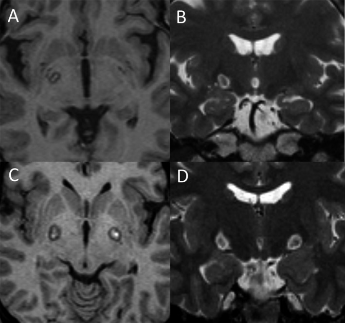
Figure 1.

MRI images of the patients after undergoing pallidotomy. (A–B) show postoperative T1‐ and T2‐weighted MRI after undergoing unilateral pallidotomy. (C–D) show postoperative T1‐ and T2‐weighted MRI after undergoing simultaneous bilateral pallidotomy.
