
Figure 2.
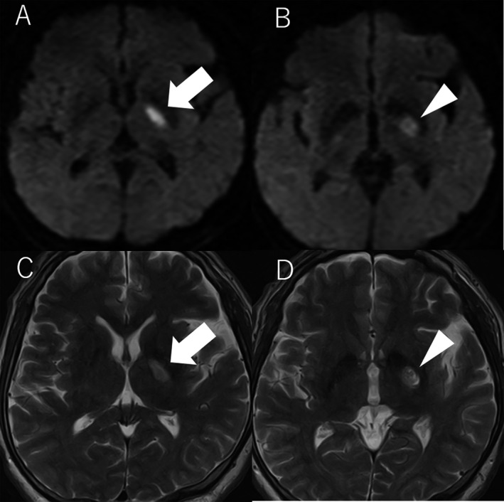
Diffusion‐weighted (A and B) and T2‐weighted (C and D) MRI images of cerebral infarction 1 month after pallidotomy. A and C show acute cerebral infarction on the left posterior limb of internal capsule (arrow). B and D show left pallidotomy lesion (arrowhead).