FIGURE 4.

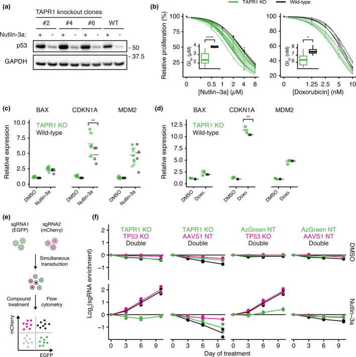
The impact of TAPR1 loss on cell fitness is TP53‐dependent. (a) NALM‐6 lysates from clonal TAPR1‐disrupted (TAPR1 knockout) or wild‐type (WT) NALM‐6 cells treated with nutlin‐3a (2 µM, 4 h) were blotted against p53 and GAPDH (1 representative blot of 3 independent replicates). (b) Relative proliferation of TAPR1‐disrupted (TAPR1 KO) or wild‐type cells treated with the indicated concentrations of nutlin‐3a or doxorubicin for 72 h. Dose–response curves were fitted and the GI50 concentration is shown as inset plots (n ≥ 3). (c) Relative expression of the indicated transcripts in wild‐type or TAPR1 KO cells treated with 2 µM nutlin‐3a or 0.1% (v/v) DMSO for 4 h (n ≥ 4). (d) Relative expression of the indicated transcripts in wild‐type or TAPR1 KO cells treated with 0.5 µM doxorubicin (Doxo.) or 0.1% (v/v) DMSO for 4 h (n ≥ 2). (e) Competitive growth assay schematic for NALM‐6 cells transduced with non‐targeting sgRNAs and sgRNAs targeting TAPR1 and TP53. (f) sgRNA enrichment in NALM‐6 cells treated with 2 µM nutlin‐3a or 0.1% (v/v) DMSO shown for the indicated TAPR1/TP53 sgRNA combinations (n = 2)
