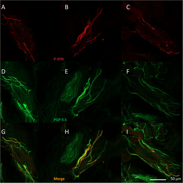
Figure 2.

Sample images of cutaneous phosphorylated alpha‐synuclein. (A–I) Examples from three different patients with POTS of a blood vessel with surrounding vasomotor nerve fibers with phosphorylated alpha‐synuclein shown in red (A–C) and vasomotor nerve fibers shown in green stained with protein gene product 9.5 (D–F), overlapping merged images in (G–I).
