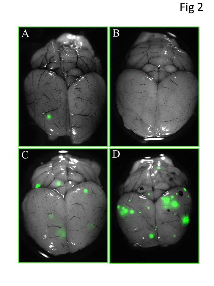
Figure 2. Images of brain metastases from PCI and treatment groups at 8 weeks after injection of GFP-labeled tumor cells.

Brain images were obtained with a fluorescent stereomicroscope. Panels A and B show representative images with (A) and without (B) brain metastases after receiving 4 Gy of whole-brain irradiation 5 days after tumor-cell injection; panel C, image from a mouse that received 4 Gy whole-brain irradiation at 3 weeks after tumor-cell injection; and panel D, image from a mouse that received 4 Gy of whole-brain irradiation 6 weeks after tumor-cell injection. Metastatic foci were the smallest in the mice irradiated 5 days after tumor-cell injection.
