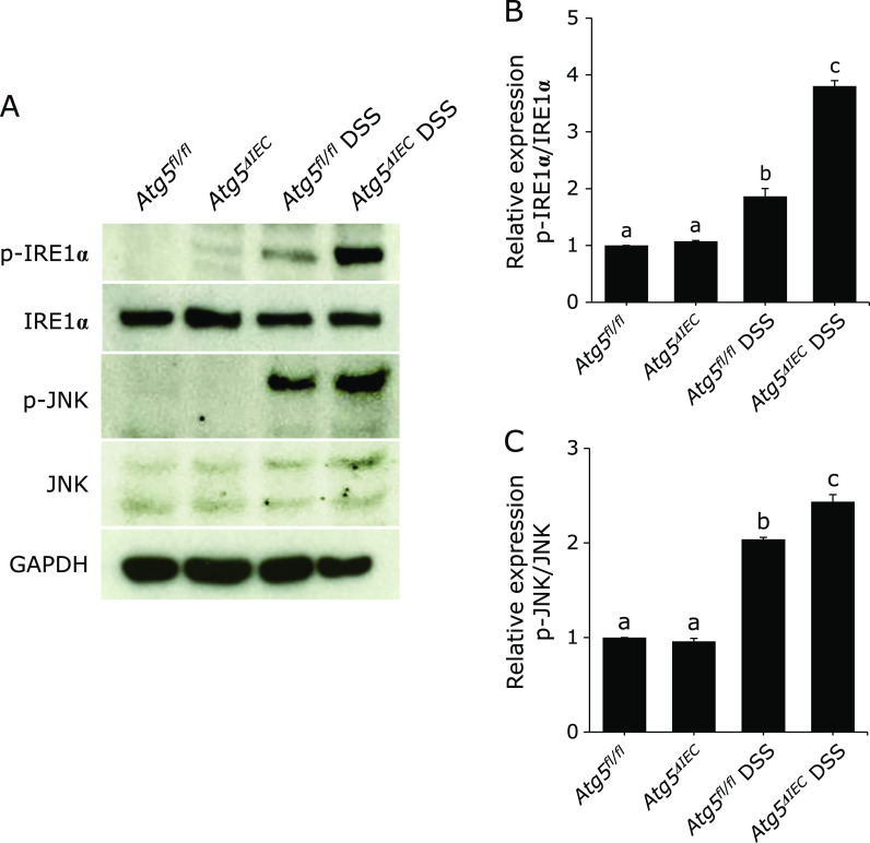
Fig. 5.
Immunoblot for phosphorylated (p)-IRE1α, IRE1α, p-JNK and JNK in the isolated colonic epithelial cells. (A) Total protein was isolated from colonic epithelial cells. GAPDH was used as a loading control. The pictures are representative of four independent experiments. Relative expression of p-IRE1α (B) and p-JNK (C). The data are expressed as means ± SEM (n = 4 mice/group). Values not sharing a letter are significantly different (p<0.05).

