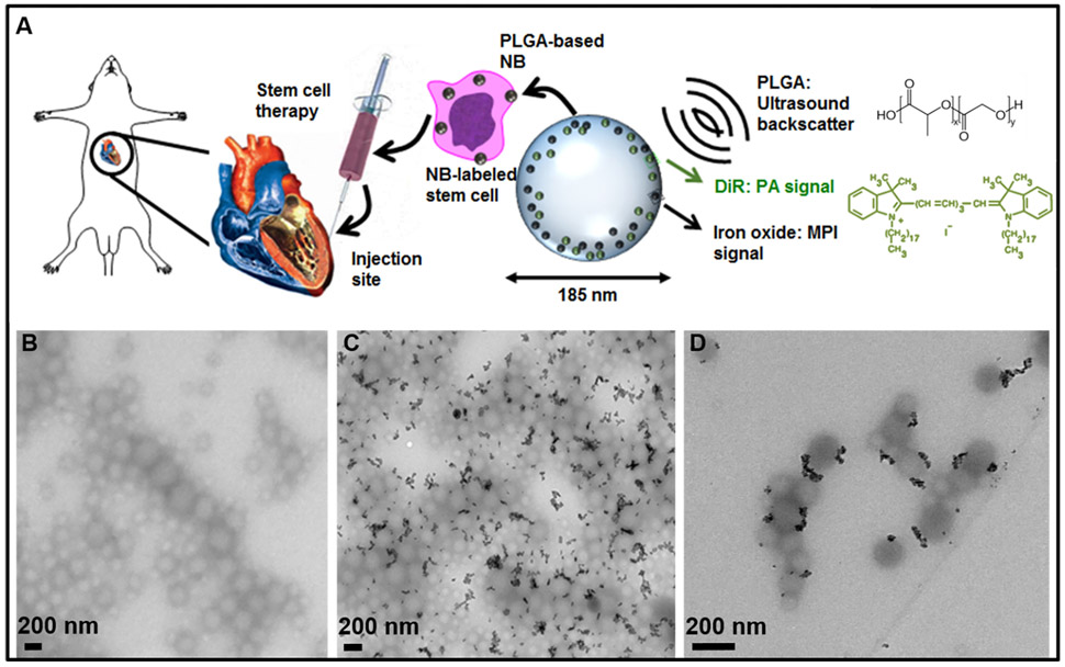
Figure 1.
Nanobubble schematic and synthesis. (A) Schematic of cells labeled with the trimodal nanobubble being injected intracardially into a murine model. The PLGA and DiR molecular structures are shown. (B) TEM image of PLGA nanobubbles without iron oxide. (C) TEM image of iron oxide-loaded PLGA particles. (D) Higher-magnification TEM image of iron oxide-loaded PLGA particles.

