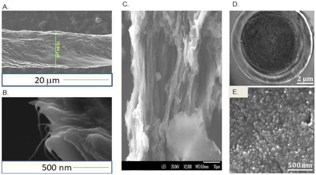
Figure 1: SEM Images of Carbon Nanotube Fibers and Yarn.

(A). SEM Image of a PEI CNT fiber with darker regions containing more conductive CNTs. (B) SEM image of a PEI CNT fiber end. Thin whiskers of individual CNTs protrude from the bundles in the cross-section. (C). SEM of Acid Spun CNT fiber. (D) A beveled end a CNT yarn microelectrode. (E) High magnification CNT yarn with 30-50 nm diameter CNTs bundled tightly together to form a nanostructured surface. Panels D and E are taken from reference (32).
