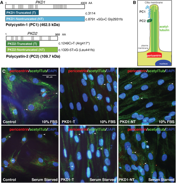
Fig 1.

Primary cilia visualized in preosteoblasts derived from patients with autosomal dominant polycystic kidney disease (ADPKD). (A) Diagram of PKD1 and PKD2 genes encoding polycystin‐1 (PC‐1; 462.5 kDa) and polycystin‐2 (PC‐2; 109.7 kDa), respectively. The patients with ADPKD chosen were identified to contain truncating or nontruncating variations of both genes. PKD1 mutations include truncation (navy) at codon 3114 or a nontruncated form (aqua) with mutations at codon 8791. PKD2 mutations include truncation (dark green) at codon 1249 and nontruncated (light green) at codon 1320. (B) Cartoon depiction of primary cilia including PC‐1 and PC‐2 in the primary cilia, acetylated tubulin (green), and the basal body with pericentrin (red) at the base of the cilium. (C) Primary cilia (arrows) marked by pericentrin (red) and acetylated tubulin (green) in primary cultured osteoblasts from patients with ADPKD (PKD1) and healthy controls in 10% serum or serum‐starved conditions (24 hours), assessed by immunofluorescence staining; the nuclei were marked by DAPI (blue). Scale bar represents 50 μm. acetylTub = acetylated‐α‐tubulin; DAPI = 4,6‐diamidino‐2‐phenylindole.
