
Fig 3.
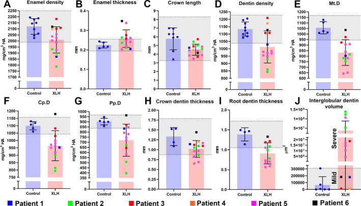
Mineralization defects associated with X‐linked hypophosphatemia rickets (XLH) in primary teeth. (A‐I) Individual values, means, and SDs from μCT analyses of dental tissues of patients with XLH versus control individuals. Control 95% CIs are shaded in gray. Patients 1–6 are color coded to recognize patterns in data across measurements. Mt.D = Mantle dentin (outermost); Cp.D = circumpulpal dentin (middle); Pp.D = proximal pulpal dentin (inner and last formed). (J) Measured volumes of interglobular dentin. Values for mildly affected patients with XLH fall within the 95% CI, and values for severely affected patients are substantially elevated above the 95% CI.