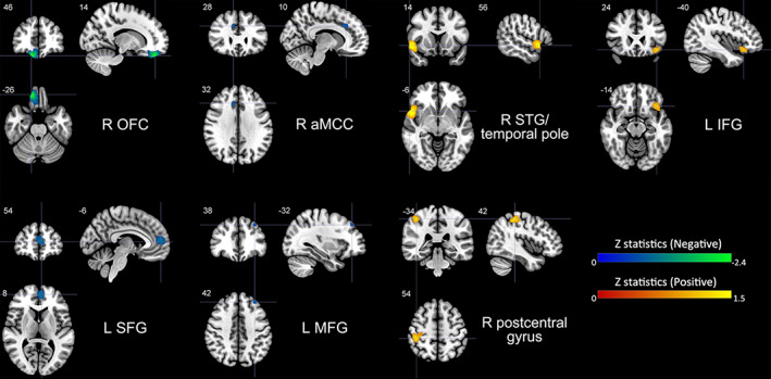
FIGURE 2.

Brain regions where the volumetric GM alterations linked with trait impulsivity. Clusters were exhibited in the sagittal, axial, and coronal planes at voxel‐wise p < .005, z > 1, and cluster size >10 voxels. Regions with negative correlates were shown in blue or green and positive correlates in red or yellow. GM, gray matter; L, left; R, right; OFC, orbitofrontal cortex; SFG, superior frontal gyrus; aMCC, anterior midcingulate cortex; MFG, middle frontal gyrus; STG, superior temporal gyrus; IFG, inferior frontal gyrus
